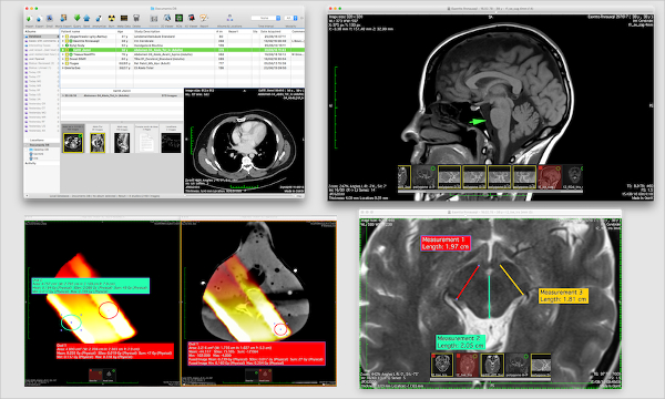

CTA examinations of patients with suspected carotid artery stenoses were used for the evaluation of the software assistant. It should enable the visualization of the vessel lumen and the quantitative evaluation of a stenosis. Purpose: A software assistant for automatic evaluation of CT-angiograms (CTA) was developed. Software-assisted CT-postprocessing of the carotid arteries A dental CT software program is an improved imaging modality for assessing jaw cysts and findings which tend to indicate odontogenic keratocysts are marginal scalloping, mandibular ramus involvement, prominent spread along the marrow space and multilocularity The findings of the other 12 cysts (eight maxillae and four mandibles) were unilocularity in all, smooth inner margin in ten, height to length ratio below 60% in only two, and ramus involvement in none. Dental CT findings of five mandibular odontogenic keratocysts were scalloped margin in all, mandibular ramus involvement in four, height to length ratio below 60% in four ,and multilocularity in two. Images of jaw cysts obtained with the dental CT software program delineated much more clearly than conventional images the status of neurovascular bundle and cortical bone, but there was no clear difference between the two modalities in delineating tooth root erosion. Seventeen lesions icomprised 15 odontogenic cysts (five odontogenic keratocysts, five radicular, three residual and two dentigerous cysts) and two non-odontogenic cysts (one nasopalatine duct cyst and one postoperative maxillary cyst). For the delineation of involvement or displace-ment of neurovascular bundles, cortical erosion, perforation or expansion, and tooth root resorption by the jaw cysts, images from this program were compared to conventional images. Seventeen patients with proven jaw cysts(8 maxillae and 9 mandibles) were evaluated with a dental CT sofware program for location, locularity, the presence or absence of marginal scallping, and height to length ratio. To evaluate the usefulness of a dental CT software program in the assessment of jaw cysts and in the differentiation of odontogenic keratocysts and other cysts. Lee, Jung Man Shin, Sang Hoon Lee, Won Hoon Oh, Kyu Hyen Jung, Hak Young Lee, Young Hwan Sung, Nak Kwan Jung, Duck Soo Kim, Ok Dong Imaging of the jaw cysts with a dental CT software program : distinction of odontogenic keratocysts from other cysts

Therefore, dental CT software program can play an important role in the preoperative assessment of mandible and maxilla for dental implants and other surgical conditions Reformatted images using dental CT software program provided excellent delineation of the jaw anatomy. In maxilla, anatomy related to neurovascular bundle(greater palatine foramen and groove, nasopalatine canal and incisive foramen) and other anatomy(alveolar process, maxillary sinus and nasal fossa) were also well delineated. Anatomy related to neurovascular bundle(mandibular foramen, inferior alveolar canal, mental foramen, canal for incisive artery, nutrient canal, lingual foramen and mylohyoid groove), muscular insertion(mylohyoid line, superior and inferior genial tubercle and digastric fossa) and other anatomy(submandibular fossa, sublingual fossa, contour of alveolar process, oblique line, retromolar fossa, temporal crest and retromolar triangle) were well delineated in mandible. Reformatted images obtained by the use of bone algorithm performed on GE HiSpeed Advantage CT scanner were retrospectively reviewed for detailed anatomy of jaw. We evaluated 13 mandibles and 7 maxillae of 15 subjects without bony disease who were being considered for endosseous dental implants. The purpose of this study is to describe the method of the technique and to identify the precise anatomy of jaw.

Kim, Myong Gon Seo, Kwang Hee Jung, Hak Young Sung, Nak Kwan Chung, Duk Soo Kim, Ok Dong Lee, Young Hwanĭental CT software program can provide reformatted cross-sectional and panoramic images that cannot be obtained with conventional axial and direct coronal CT scan. International Nuclear Information System (INIS) Imaging of jaw with dental CT software program: Normal Anatomy


 0 kommentar(er)
